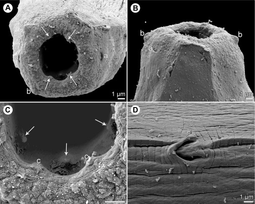
Figure 8.
Johnstonmawsonia sp. from Carangoides fulvoguttatus, scanning electron micrographs of nongravid female. A, B: Cephalic end, apical and dorsoventral views, respectively (arrows indicate sublabia). C: Detail of mouth, apical view (arrows indicate inner prostomal teeth). D: Excretory pore, ventral view. Abbreviations: a, amphid; b, submedian cephalic papilla; c, sublabium.

