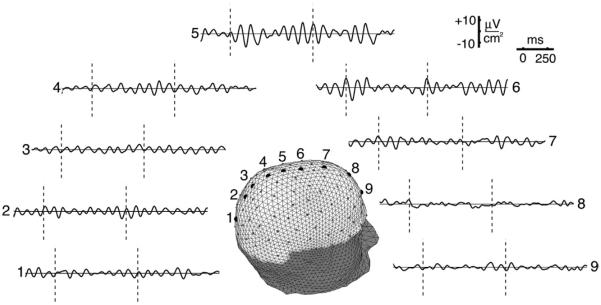
Fig. 4.
High resolution estimates (HR-EEG) of the same EEG data shown in Fig. 3 are obtained by passing the recorded waveforms from 111 sensors (excluding 17 lower edge sites for technical reasons) to the New Orleans spline-Laplacian algorithm that “knows” the basic physics of current spread through brain, skull, and scalp tissue. Nearly identical results are obtained from the independent Melbourne dura imaging algorithm, which unlike the Laplacian, is based on a 3-sphere head model. These two algorithms essentially filter out the very large scale (low spatial frequency) scalp potentials, which consist of some unknown combination of passive current spread and genuine large scale cortical source activity. The largest high resolution signals occur at middle sites 5 and 6 where the unprocessed potentials are smallest. The apparent explanation follows from Fig. 2; high resolution estimates are more sensitive to sources in smaller patches (dipole layers) of cortex. The alpha rhythms apparently have multiple contributions, including a global process (e.g., standing wave) and a local source region near the middle of the electrode array. This local oscillation, called the mu rhythm, is blocked by movement or planned movement because of its proximity to motor cortex. A second local alpha process, blocked by eye opening, was found in occipital cortex (not shown). Reproduced with permission from (Nunez and Srinivasan, 2006).

