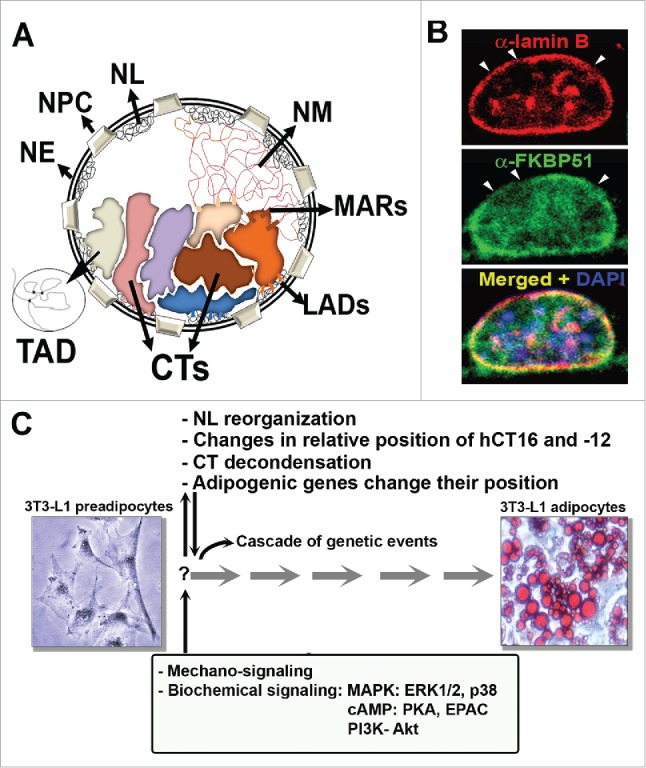
Figure 1.

Nuclear architecture during adipogenesis. (A) Schematic representation of the compartments of the nucleus in interphase: NE: nuclear envelope, NPC: nuclear pore complex, NL: nuclear lamina, NM: nuclear matrix, CTs: chromosome territories, LADs: lamin attachment domains, MARs: Matrix attachment regions; TADs: topologically associating domains. (B) Reorganization of the NL during adipocyte differentiation. 3T3L1 preadipocytes grown on coverslips were induced to differentiate for 24h. Indirect immunofluorescence and confocal microscopy imaging shows lamin B (red), FKBP51 (green) and chromatin stained with DAPI (blue), as described.98 Observe the discontinuous staining of lamin B (arrow heads) due to the reorganization of the NL. (C) Summary of the events of nuclear reorganization that were described during adipogenesis. Images depict 3T3-L1 preadipocytes and adipocytes, the latter with lipid vesicles stained with Oil Red O, as described.98
