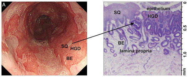
Figure 1.
Imaging of Barrett’s oesophagus (BE). (A) Wide-field imaging is needed to localise neoplastic lesions, identify tumour margins and evaluate for cancer recurrence. White light image shows patches of squamous (SQ) in BE. An area of high-grade dysplasia (HGD) is not visibly distinct. (B) Cross-sectional imaging is needed to assess depth of early cancer invasion (T1a vs T1b). Histology (H&E) shows feature of both SQ and HGD.

