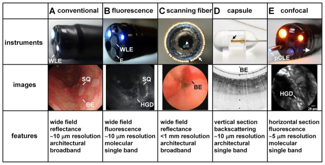
Figure 3.
Novel imaging instruments. (A) Conventional white light endoscopy(WLE) shows squamous (SQ) patches within Barrett’s oesophagus (BE). (B) Fluorescence (F) endoscope shows molecular images of high-grade dysplasia (HGD; high signal) next to SQ (low signal). (C) Scanning fibre endoscope inserted transnasally shows patches of BE. (D) Capsule endoscopy with tethered probe shows cross-sectional image of BE on optical coherence tomography (images courtesy of G. Tearney and M. Gora). (E) Confocal laser endomicroscope (CLE) passes through instrument channel of endoscope to shows optical cross-section of HGD on molecular imaging in vivo. pCLE, probe-based CLE.

