
FIG 2.
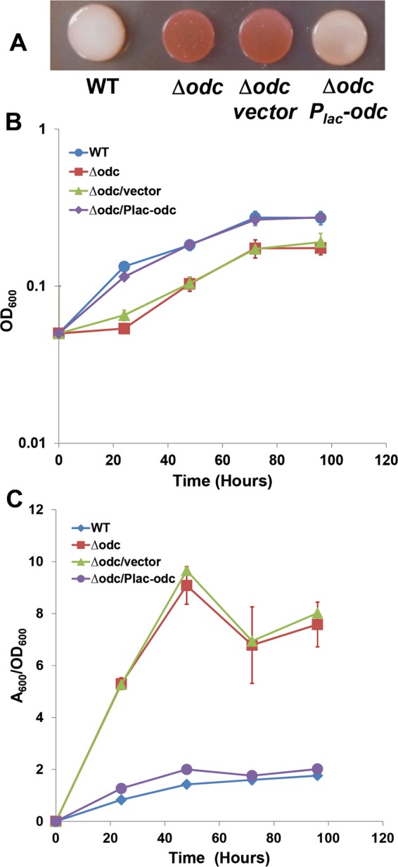
The odc mutant has elevated Congo red staining, growth deficiency, and elevated biofilm formation. (A) Congo red staining on an ATGN-CR agar plate; (B) growth curve from cultures used in the biofilm assay; (C) biofilm assays on coverslips. A. tumefaciens C58 and derivatives were incubated in 12-well plates with PVC coverslips for 0 to 96 h at 28°C. After being rinsed, coverslips were stained with 0.1% crystal violet and adherent biomass was measured as the absorbance of acetic acid-solubilized crystal violet, normalized for planktonic culture growth (A600/OD600) from the same well. Derivatives harboring the Plac-odc plasmid and the vector control were induced with 500 μM IPTG. Error bars show the standard deviation from a minimum of three biological replicates. WT, wild type.