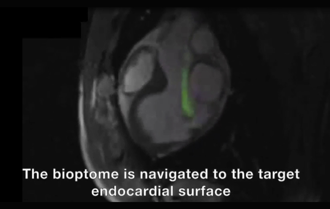
Figure 3.
Real-Time MRI-Guided Endomyocardial Biopsy
(A) Rapid frame-rate real-time magnetic resonance imaging (MRI) during navigation of the active visualization MRI bioptome within the left ventricle to the target endocardial surface. The jaws appear as a passive artifact (arrow). (B) After systemic gadolinium contrast administration, the lesion is visible using inversion-recovery real-time MRI (arrows). Real-time MRI-guided biopsy specimens viewed under (C) transmitted light and (D) ultraviolet light. X-ray fluoroscopy–guided biopsy specimens viewed under (E) transmitted light and (F) ultraviolet light. Also see Video 1.
A demonstration of real-time MRI guided endomyocardial biopsy in an animal model.


