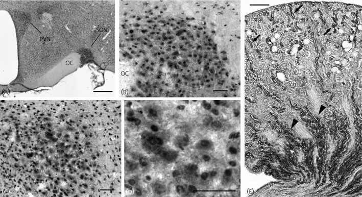
Figure 3.

Immunocytochemical localisation of 11β‐hydroxysteroid dehydrogenase type 2 (11β‐HSD2) in sections of the hypothalamus and kidney from a Wistar–Kyoto (WKY) rat. (a) 11β‐HSD2 immunoreactivity was observed in essentially all neuronal nuclei in the brain section. This ubiquitous nuclear labelling was most likely nonspecific labelling; however, the immunoreactivity was dense in the paraventricular nucleus (PVN) and supraoptic nucleus (SON). (b) In the SON, magnocellular neurosecretory cells (MNCs) containing cytoplasmic immunoreactivity were observed. Note that cells outside of the SON are devoid of cytoplasmic immunoreactivity. (c) In the PVN, the cells possessing cytoplasmic immunoreactivity were mostly located in the posterior magnocellular cluster. (d) A higher power view demonstrates cytoplasmic immunoreactivity in MNCs in the PVN. (e) Immunocytochemical localisation of 11β‐HSD2 was also performed in the kidney. In the kidney cortex, an intense 11β‐HSD2 immunoreactivity was present in the tubules for which the cellular morphology and location resemble that of the distal tubules (arrows). In the kidney medulla, a strong 11β‐HSD2 immunoreactivity was present in collecting ducts (arrowheads). OC, optic chiasm. Scale bar = 500 μm in (a) and (e), 50 μm in (b–d).
