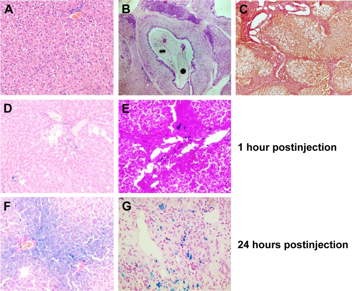
Figure 8.
Histological analysis of the liver microsection.
Notes: Hematoxylin and eosin staining of (A) healthy (×100) and (B) O. felineus-infected hamster liver (×50). (C) Van Gieson staining of O. felineus-infected hamster liver (×100). Prussian blue staining of (D, F) healthy (×200) and (E, G) O. felineus-infected hamster liver (×200) harvested 1 and 24 hours after the administration of MNPs-NH2 at a dose of 0.6 mg kg−1.
Abbreviations: O. felineus, Opisthorchis felineus; MNPs, magnetic nanoparticles.

