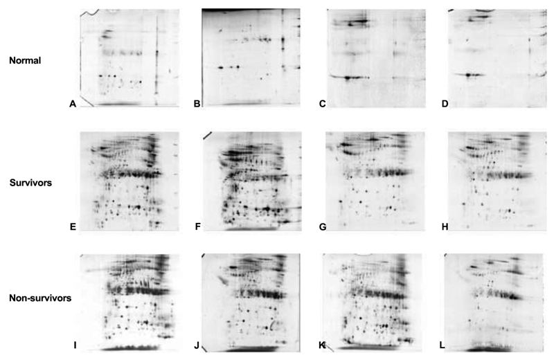
Figure 2.
Cerebrospinal fluid was separated using 2-dimensional polyacrylamide gel electrophoresis. The gels were grouped into the following 3 groups: normal (controls), survivors, and nonsurvivors. Two-dimensional polyacrylamide gel electrophoresis separates proteins on the basis of isoelectric point and molecular weight. The proteins are visualized using silver stain and analyzed for differences. It can be observed that cerebrospinal fluid of controls has less protein, compared with that of the survivors and nonsurvivors. It can also be observed that the determination of differences between survivors and nonsurvivors is complex.

