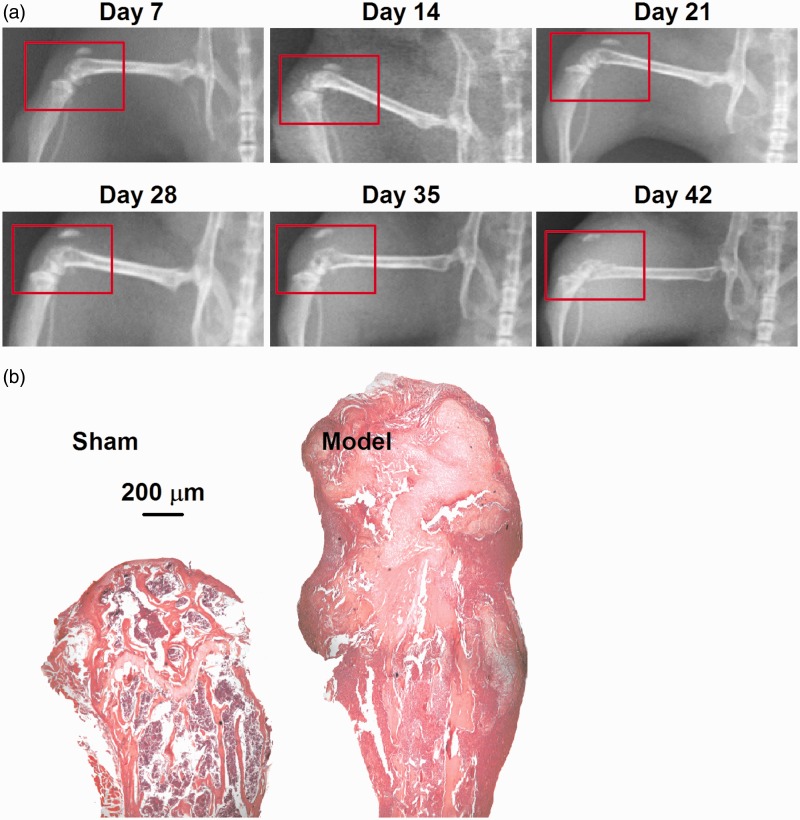
Figure 1.
Establishment of bone cancer model. (a) X-ray images of the model femur showed the progressive loss of mineralized bone after the injection of sarcoma cells. Numbers represent days after surgery. Red circles indicate the operative side. (b) H&E staining showed the pathological structure of femora. In the sham bone (left), there is a clear separation of mineralized bone (normal, pink) and marrow cells (with large number of inflammatory cells infiltration, purple). In the model bone (right), the smaller and more densely packed cancer cells (purple) have largely replaced the marrow cells and destructed the mineralized bone to fracture (pink) in the intramedullary space.

