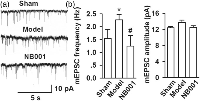
Figure 5.
Miniature EPSCs in ACC neurons. (a) Representative mEPSCs recorded in pyramidal neurons at a holding potential of −70 mV from sham (saline), model (saline), and NB001 (30 mg/kg, ip, twice per day for three days) mice. (b) Cumulative frequency (left) and amplitude (right) histogram of the mEPSCs in neurons from sham, model, and NB001 mice. Values were obtained by normalizing the mean peak currents at each holding potential to a holding potential of −70 mV. Inset shows the mEPSCs recording at +50 and −70 mV. Data are presented as mean ± SE, *p < 0.05 versus sham, #p < 0.05 versus model.

