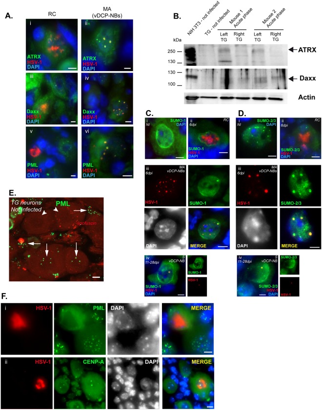
Fig 2. HSV-1 MA pattern corresponds to vDCP-NBs and contains SUMO proteins.
(A) Immuno-DNA-FISH showing HSV-1 genomes (red), promyelocytic leukemia nuclear bodies (PML-NB)–associated proteins (green), and cellular chromatin (DAPI, blue). (B) WB of ATRX and Daxx in TG of uninfected or infected mice during acute infection. Infected (left TG) and not-infected (right TG) TGs of the same mouse (two mice) were harvested 6 dpi, and treated to perform WB. Actin is shown as a loading control. NIH3T3 is shown as a cellular control. (C) and (D) Immuno-DNA-FISH showing HSV-1 genomes (red), small ubiquitin modifier (SUMO) proteins (green), and cellular chromatin (DAPI, blue/grey). (C) SUMO-1 and (D) SUMO-2/3 detection in (i) non-infected neurons, (ii) RC-containing neurons and (iii, iv) MA/vDCP-NBs or S/vDCP-NB-containing neurons. (E) IF for detection of PML (green) in uninfected TG neurons and satellite cells. Arrows point out PML-NBs in neurons or satellite cells, arrowheads point out neurons without PML-NBs. (F) Immuno-DNA-FISH showing HSV-1 genomes (red, RC) and (i) PML or (ii) CENP-A (green), in neurons. DAPI staining (grey) shows nuclei. Scale bars represent 10 μm.

