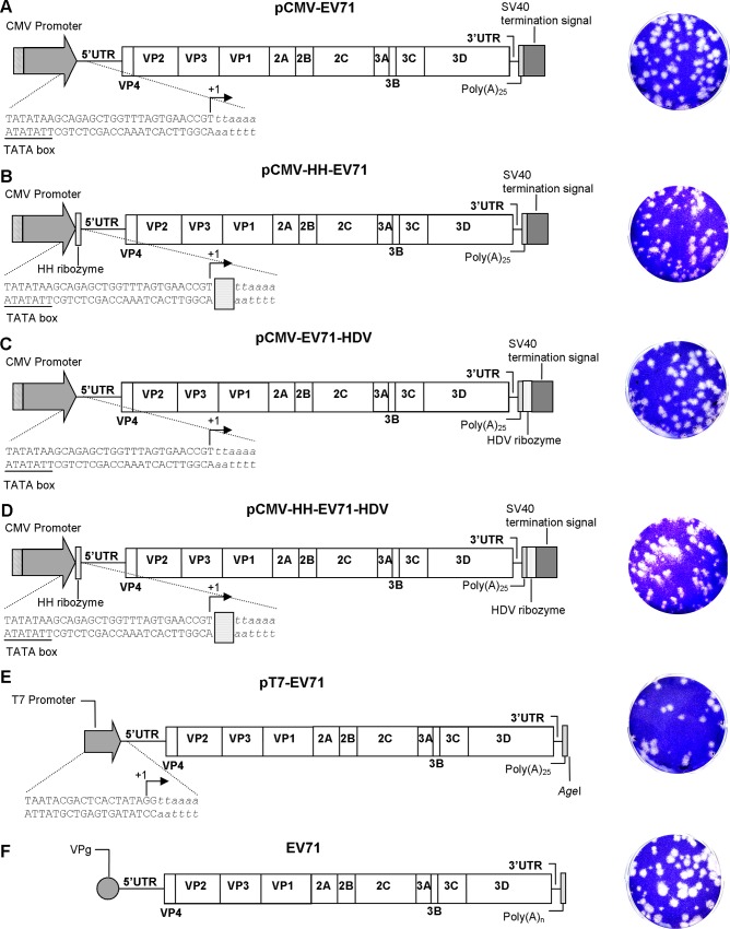
Fig 1. Schematic illustrations of CMV promoter-driven and T7 promoter-driven EV A71 infectious cDNA clones.
The schematic illustrations of (A) pCMV-EV71, the CMV promoter-driven EV-A71 infectious clone; (B) pCMV-HH-EV71, the CMV promoter-driven EV-A71 infectious clone with the HH ribozyme before the 5’UTR; (C) pCMV-EV71-HDV, the CMV promoter-driven EV-A71 infectious clone with the HDV ribozyme after the EV-A71 poly(A)25 tail; (D) pCMV-HH-EV71-HDV, the CMV promoter-driven EV-A71 infectious clone with the HH ribozyme upstream of EV-A71 5’UTR and HDV ribozyme downstream of EV-A71 poly(A)25; (E) pT7-EV71, the T7 promoter-driven EV-A71 infectious clone; and (F) wild-type EV-A71. Italicized nucleotides indicate EV-A71 genomic DNA. Arrows indicate transcription start sites. The plaque morphologies of each clone-derived EV-A71 are shown in the right panel.

