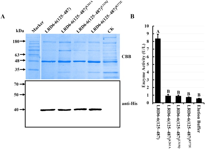
Fig 4. In vitro ATPase activity determination of LRD6-6.
(A) Detection of the N-terminal truncated recombinant protein His–LRD6-6(125–487), and its variants, His–LRD6-6(125–487)K261A, His–LRD6-6(125–487)E315Q and His–LRD6-6(125–487)R372E, purified from E. coli by coomassie brilliant blue staining (upper panel) and western blot with anti-His (lower panel). (B) In vitro ATPase assay on recombinant proteins His–LRD6-6(125–487), His–LRD6-6(125–487)K261A, His–LRD6-6(125–487)E315Q and His–LRD6-6(125–487)R372E. ATPase activities were measured using a malachite green-based colorimetric approach. The ATPase activities with mean values ± SEM of four replications were shown. Statistical significance comparison was conducted with ANOVA (P < = 0.01), where different capital letters above columns indicate significant differences, whereas the same letter indicates no significant differences.

