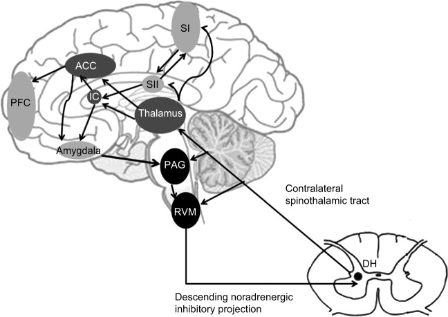
Figure 1.
A schematic representation of pain modularity circuitry and the pain matrix.
Notes: Nociceptive inputs enter the spinal DH through primary afferent fibers that synapse onto transmission neurons. The projection fibers ascend through the contralateral spinothalamic tract targeting the thalamus, and collateral projections also target mesencephalic nuclei, including the RVM and the midbrain PAG. In the pain matrix, there are two complementary pathways through which pain processing takes place. The medial pathway (dark gray) projects from the medial thalamus to the ACC and IC and processes the affective-motivational component of pain (ie, unpleasantness). The lateral pathway (light gray) projects from the lateral thalamus to the primary and secondary somatosensory cortices (SI and SII) and IC and processes the sensory-discriminative aspect of pain (ie, location and intensity). Increased activation of the PFC is related to decreased pain affect purportedly by inhibiting the functional connectivity between the medial thalamus and the midbrain. Descending projections from the hypothalamus (not shown), amygdala, and rACC feed to the midbrain PAG and to the medulla. Neurons within the RVM project to the spinal or medullary DH to inhibit pain experience. © Springer Science+Business Media, LLC 2012. Reproduced with permission of Springer. Jones AK, Huneke NT, Lloyd DM, Brown CA, Watson A. Role of functional brain imaging in understanding rheumatic pain. Curr Rheumatol Rep. 2012;14(6):557–567.36
Abbreviations: DH, dorsal horn; RVM, rostral ventral medulla; PAG, periaqueductal gray; ACC, anterior cingulate cortex; IC, insular cortex; PFC, prefrontal cortex; rACC, rostral anterior cingulate cortex.

