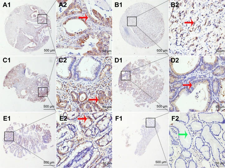
Figure 1.
PHGDH protein expression in benign and malignant gastric tissue samples in TMA sections.
Notes: (A1 and A2) Well-differentiated gastric cancer tissue with moderate PHGDH expression; (B1 and B2) poorly differentiated gastric cancer tissue with strong PHGDH expression; (C1 and C2) High-grade intraepithelial neoplasia with moderate PHGDH expression; (D1 and D2) Low-grade intraepithelial neoplasia with weak PHGDH expression; (E1 and E2) Intestinal metaplasia with weak PHGDH expression; (F1 and F2) Normal surgical margin of gastric cancer, negative. Original magnification ×40 (bar =500 μm) in (A1), (B1), (C1), (D1), (E1), and (F1) and ×400 (bar =50 μm) in (A2), (B2), (C2), (D2), (E2), and (F2). Red arrows indicate positive PHGDH staining while green arrows indicate negative PHGDH staining.
Abbreviations: PHGDH, phosphoglycerate dehydrogenase; TMA, tissue microarray.

