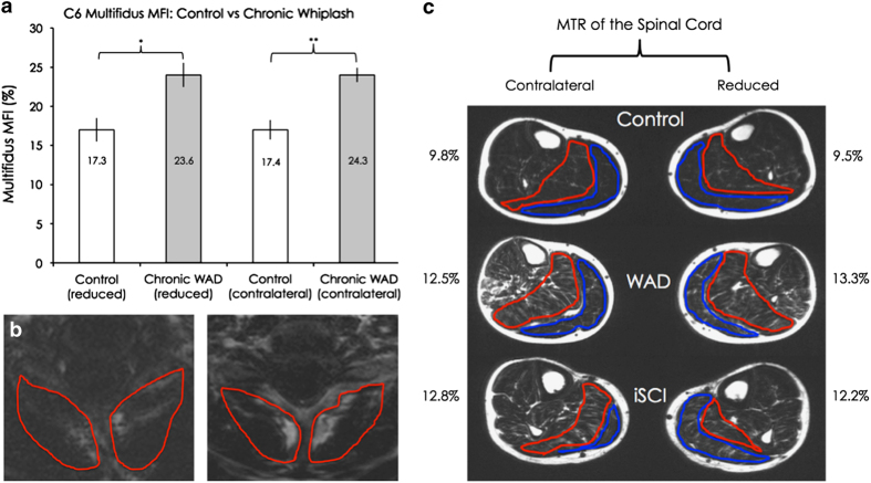
Figure 2.
(a) Group means and s.e.m. of the multifidus-semispinalis cervicus MFI on the reduced and contralateral sides, for the control and chronic WAD groups. A significant difference was found on both the reduced (P=0.02) and contralateral sides (P<0.01). (b) Fat-saturated images of the multifidus-semispinalis cervicus MFI regions of interest, at the C6 level, for one uninjured matched-control participant (left) and a participant with chronic WAD (right). (c) Group means of the plantarflexor MFI on both the reduced and contralateral sides, for all three groups. Differences between the control and chronic WAD mean values were non-significant on the reduced side following Bonferroni correction (P=0.04), while the other comparisons were not significant (P range: 0.15–0.85).

