. 2016 Aug 15;5:e15200. doi: 10.7554/eLife.15200

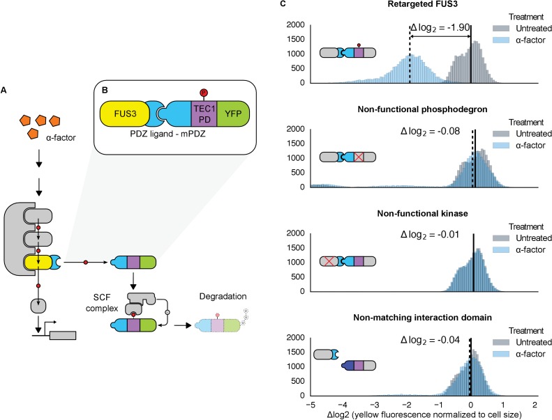
Figure 1. Rewiring the mating cascade MAPK, Fus3, to regulate the degradation of YFP.

(A) The core components of the yeast mating cascade. The yeast mating factor – α-factor – triggers the sequential activation of the kinases Ste11 and Ste7 (rounded gray rectangles) followed by the MAPK, Fus3 (yellow). Arrows with red circles denote phosphorylation-mediated regulation. All three kinases are organized on the scaffold Ste5 (also gray). Among other effectors, Fus3 activates the transcription factor Ste12 (rounded gray box). (B) Fus3 targeted regulation of YFP (green). The colocalization was controlled by the addition of the mPDZ domain to YFP and a PDZ ligand to Fus3 (light blue). Degradation was mediated by the addition of a phosphodegron derived from the transcription factor Tec1 (purple). Upon activation of the mating pathway, Fus3 phosphorylates the phosphodegron fused to YFP, resulting in the recruitment of an E3 ubiquitin ligase and the ubiquitination and subsequent degradation of YFP. (C) Cells bearing the modified Fus3 and either the fully functional system, a reporter construct with an inactivated phosphodegron, a Fus3 with its kinase activity knocked out or an unmatched interaction domain (an SH3 domain instead of mPDZ) were grown to log phase and induced with 10 μM α-factor (blue histograms) or un-induced (gray histograms). Data shown are from 3 hrs post-induction. The vertical dashed black lines on the histograms represent medians of treated populations and solid black lines represent medians of untreated populations. In all figures, the fluorescence has been normalized to the cell size (see Figure 1—figure supplement 1). Full time-course experiments appear in the supplement to Figure 2.

DOI: http://dx.doi.org/10.7554/eLife.15200.003

Figure 1.

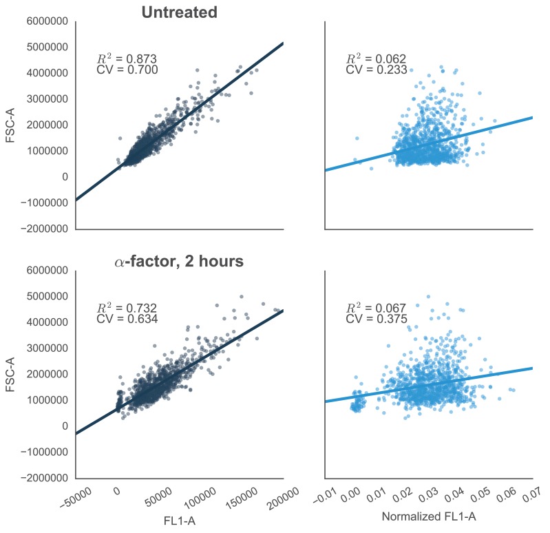
Figure 1—figure supplement 1. Reducing the variability of single-cell fluorescence by accounting for cell-to-cell variation in cell size.

Figure 1—figure supplement 1.

In yeast constitutively expressing a single-copy fluorescent protein inserted in the genome, fluorescence (FL1-A) is strongly correlated with cell size (approximated by FSC-A)—shown by R2 values. This is true in both cells that are untreated (top left) or treated with 10 µM α-factor (bottom left). The effect is likely due to the way that flow cytometers measure fluorescence, where cells with the same concentration of fluorescent protein – but with different volumes – will have different levels of fluorescence. For example, smaller cells that have just divided will have a lower fluorescence value than larger cells that are just about to divide. Normalizing by cells size – dividing FL1-A by FSC-A – reduces the coefficient of variation (CV) of the fluorescent signal by ~67% or ~41% in untreated or treated cells, respectively (graphs on right).
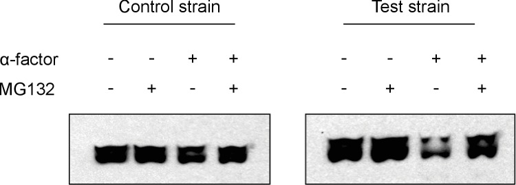
Figure 1—figure supplement 2. Western Analyses of degradation assays.

Figure 1—figure supplement 2.

The representative western blots above show results from degradation assays on a test strain with the entirely functional system described in Figure 1 and a control strain that has a non-functional phosphodegron. We observe that upon treatment with α-factor, the test strain has a significantly fainter band as compared to the untreated lane, whereas the control strain does not. This is consistent with our flow cytometry observations. We also observe that when cells are treated with MG132, a proteasome inhibitor, the alpha-factor triggered degradation in the test strain is prevented, implying that the degradation we observe is indeed due to the proteasome as hypothesized.
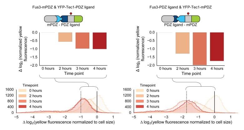
Figure 1—figure supplement 3. Swapping interaction domains between kinase and substrate.

Figure 1—figure supplement 3.

Population histograms and medians of YFP fluorescence signal normalized by cell size for yeast strains that have either a Fus3 kinase fused to a PDZ ligand and YFP fused to a PDZ domain (right) or Fus3 kinase fused to a PDZ domain and YFP fused to a PDZ ligand. These strains were diluted down from saturated overnights and were grown for 5 hrs to log phase and then cytometry reads were performed after 2, 3 and 4 hrs post induction with α-factor. The dashed black lines represent the median fluorescence at 4 hrs and the solid black lines are the media fluorescence at 0 hr.
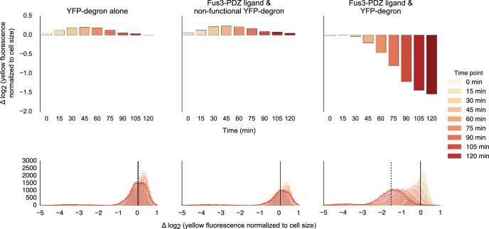
Figure 1—figure supplement 4. Fusing interaction domain to the native copy of the kinase.

Figure 1—figure supplement 4.

Population histograms (bottom) and corresponding medians (top) of YFP fluorescence signal normalized by cell size for yeast strains where the native copy of the kinase is fused to a PDZ ligand. The left most panel describes a strain with just the YFP substrate, the middle one represents a strain where the endogenous copy of Fus3 had an interaction domain fused to it but the YFP substrate has a non-functional degron and the rightmost panel describes a strain that has both an interaction domain on the Fus3 and a functional degron on the YFP substrate. These strains were diluted down from saturated overnights and were grown for 5 hrs to log phase and then cytometry reads were performed at 15 min intervals post induction with α-factor.