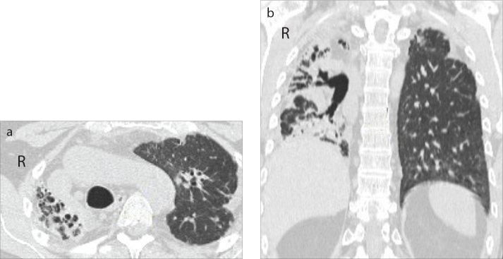
Figure 1.
a, b. Case 5: PPFE secondary to an allogeneic bone marrow transplant with asymmetric distribution on the right and left lungs. Axial (a) and coronal (b) CT images show cystic bronchiectasis, pleuroparenchymal and blotchy opacities involving the entire lung including the lower zone, together with the superior hilar retraction and the volume reduction of the entire right lung (grade 4). In the left lung, axial (a) and coronal (b) CT images show cylindrical bronchiectasis, pleuroparenchymal opacities distributed in the upper zones, without consistent volume reduction (grade 1).

