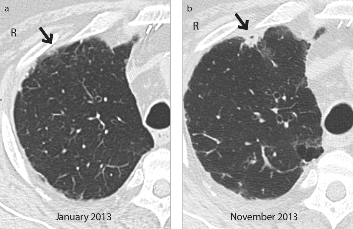
Figure 2.
a, b. Case 3: PPFE (grade 2) secondary to a double-lung transplantation. Axial CT image (a) shows subpleural ground glass opacity (arrow) in the anterior segment of the right upper lobe; it rapidly progressed into a consolidation, as shown in the axial CT scan (b, arrow) taken after nine months. Also note new reticular abnormalities and ground glass opacity in the posterior segment.

