Figure 1.
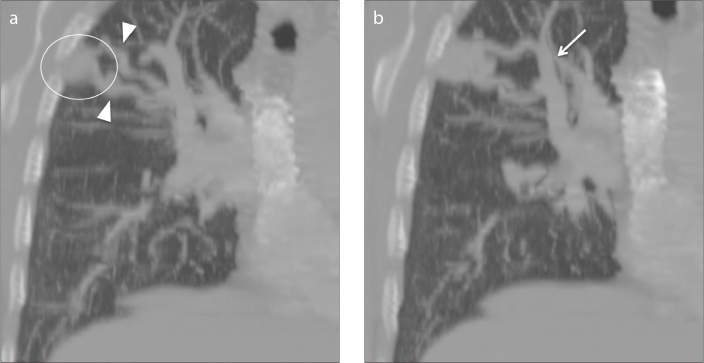
a, b. Coronal multiplanar reconstruction CT angiography images. Panel (a) shows pulmonary arteriovenous malformation (PAVM) in the upper right lobe (circle) appearing as a rounded well-circumscribed lesion with two branching afferent feeding vessels (arrowheads). Panel (b) shows a dilated efferent draining vessel (arrow).

