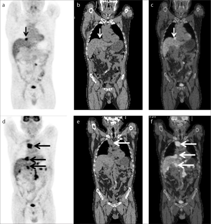
Figure 3.
a–f. A 48-year-old Chinese male presenting with fever of unknown origin. The medical history included diffuse large B-cell lymphoma. According to medical history and histopathology, the patient was diagnosed with secondary HLH associated with diffuse large B-cell lymphoma. The first coronal PET (a), CT (b) and fused (c) images showed significantly hypermetabolic lymph nodes in the retroperitoneal region (small arrows in a–c). The second coronal PET (d), CT (e) and fused (f) images showed multiple, significantly hypermetabolic lymph nodes in the thoracic, abdominal, and retroperitoneal regions (large arrows in d–f), which indicated progression of diffuse large B-cell lymphoma.

