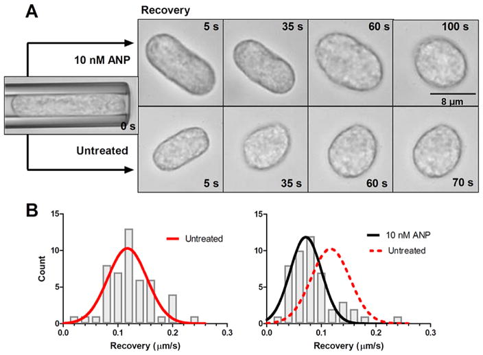
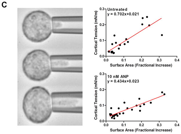
Fig. 3.
Rheological analysis of PMN cortical tension and viscosity. Untreated and 10 nM ANP treated PMN were suspended in HEPES buffered saline with 10% autologous serum to maintain unactivated and non-adherent state. (A) PMN were aspirated into the micropipette and held for 10 s prior to expulsion into the chamber. Kinetics of recovery back to spherical state (e.g. defined as length to width ratio of 1.1) was recorded and depicted in representative images. ANP treated PMN recovered ~30% slower than vehicle control. (B) Time constant for recovery was fit to a viscoelastic model yielding an estimate of cortical tension/viscosity [26,27]. Histograms of vehicle control versus ANP were Gaussian fit as depicted. Untreated PMNs (red line) versus 10 nM ANP (black) show a significant left shift in the tension/viscosity ratio due to ANP treatment (n = 50 PMN per condition analyzed from n = 4 separate donors). (C) PMN were partially aspirated into a micropipette while internal pressure was slowly increased and plotted versus protrusion length of hemispherical cap as depicted in images. Laplace’s law was used to estimate cortical tension and correlate it with a fractional increase in surface area from resting spherical state (see Methods). A linear regression was used to compute resting cortical tension in vehicle control (0.021 ± 0.013) and 10 nM ANP treated cells (0.023 ± 0.007) (n = 50 PMN analyzed from 4 separate experiments). No significance found between control and ANP treatment. (Colors are visible in the online version of the article; http://dx.doi.org/10.3233/BIR-15067.)


