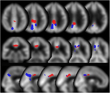
Fig. 1.

Volumes of interest (VOIs). The areas are the parts of the early Alzheimer’s disease (AD)-specific VOIs in the easy Z-score imaging system (eZIS) located in the posterior cingulate to the precunei area. Blue area: Precuneus_AD_VOIs which are the areas of overlap of the precuneus VOIs in the Automated Anatomical Labeling (AAL) atlas and the early-AD-specific VOIs. Red area: Posterior_Cingulate_AD_VOIs which are the areas of overlap of the cingulum VOIs in the AAL atlas and the early-AD-specific VOIs
