Figure 6.

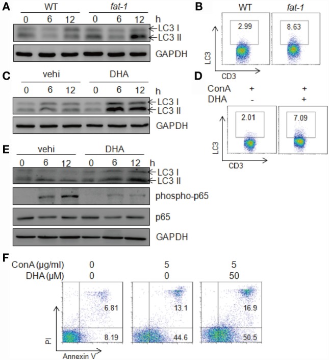
n-3 PUFAs increase autophagy levels in hepatic T cells stimulated with Con A in vitro. (A) Autophagy-associated protein LC3 in Con A (5 μg/ml)-stimulated hepatic MNCs from WT and fat-1 transgenic mice were analyzed by western blotting analysis at indicated time. (B) The frequency of LC3-producing hepatic T cells was assessed at 12 h after Con A stimulation by intercellular staining and then FACS analysis. (C) Autophagy-associated protein LC3 in Con A (5 μg/ml)-cultured hepatic MNCs in the presence or absence of DHA (50 μmol/L) by western blotting analysis. (D) The frequency of LC3-producing hepatic T cells was assessed at 12 h after Con A stimulation with or without DHA treatment by intercellular staining and then FACS analysis. Data shown represent three independent experiments with similar results. (E) Autophagy-associated protein LC3 and NF-κB p65 in Con A (5 μg/ml)-stimulated hepatic T cells with or without DHA (50 μmol/L) treatment were analyzed by western blotting analysis at indicated time. Data are representative of three independent experiments. (F) Hepatic T cells purified from WT mice were incubated with Con A (5 μg/ml) in the presence or absence of DHA (50 μmol/L) for 24 h. Cells apoptosis were determined by Annexin V/PI staining. Data shown represent three independent experiments with similar results.
