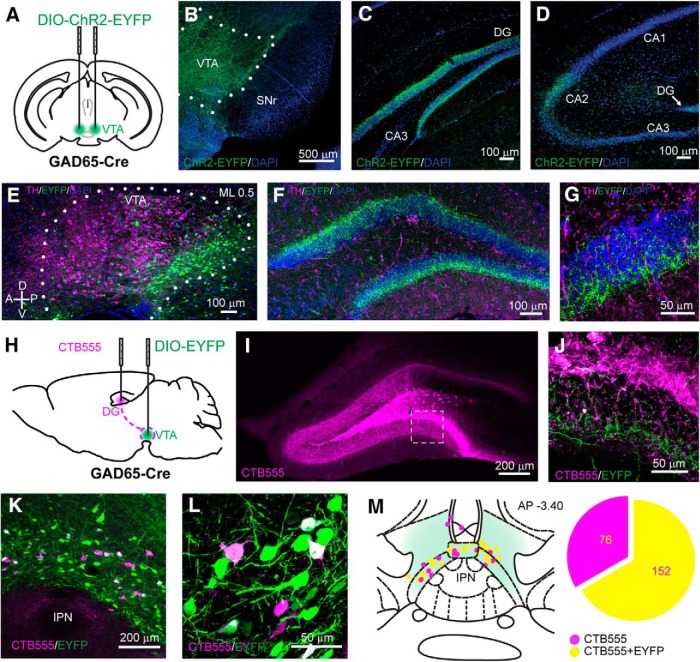
Figure 1.
VTA GABA cells project to the hippocampus. A, Schematic of the viral injection of the Cre-dependent ChR2-EYFP construct in the VTA of GAD65-Cre mice. B, Coronal section of the VTA showing the expression of ChR2-EYFP in GABA neurons. C, D, ChR2-EYFP-expressing axons are observable in the GCL of the DG (C) and in the CA2 region (D). E, Sagittal section of illustrating the anteroposterior distribution of the infected GABA neurons withing the VTA. The ML coordinate from the midline is indicated at the top. F, G, Confocal images at low (F) and high (G) magnification showing the absence of colocalization between VTA GABA fibers and TH in the DG. H, Schematic showing the injection of CTB555 into the dorsal DG and of a DIO-EYFP-expressing viral vector into the VTA of a GAD65-Cre mouse. I, Coronal section of the dDG showing the CTB555 injection site. J, Higher-magnification image detailing the presence of EYFP-expressing axons in the GCL. K, L, Confocal images at low (K) and high (L) magnification showing the retrograde labeling of CTB555 in medial VTA neurons, some of which are EYFP-expressing GABA neurons. M, Schematic representation of the localization of the retrogradely labeled neurons within the VTA and the proportion of neurons colocalizing with EYFP. The AP coordinate from bregma is indicated at the top.

