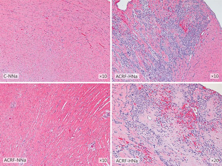
Fig. 4.
LV histology appeared normal in pair-fed controls (C-NNa) and in rats with ACRF on normal (0.6%) NaCl diets (ACRF-NNa). In ACRF rats that had received 2 weeks of high-NaCl (4%) chow (ACRF-HNa), the left ventricle showed focal areas with inflammatory cell infiltration, fibrosis, necrotic cardiomyocytes, and perivascular erythrocytes, indicating hemorrhages. Sections were stained with hematoxylin and eosin.

