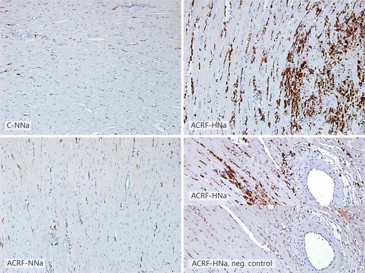
Fig. 5.
Immunohistochemistry identifying CD68-positive cells (monocytes and macrophages) in the left ventricle of pair-fed controls (C) and rats with ACRF on a normal (0.6%; NNa) or high-NaCl (4%; HNa) diet. A large proportion of the cells within injured areas in the left ventricle of ACRF-HNa rats showed CD68 positivity. In the lower right panel, a negative control, in which no primary antibody was applied, has been included. ×20.

