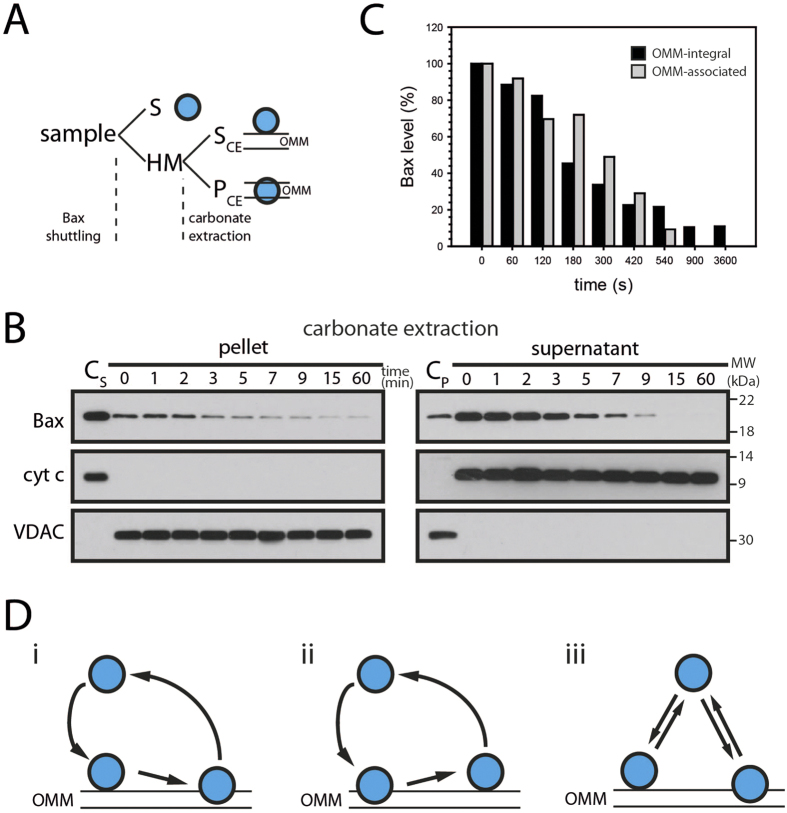
Figure 2. Membrane-integral Bax retrotranslocates.
(A) The analysis of different Bax pools after retrotranslocation involves the separation of mitochondria-containing heavy membrane fraction (HM) and supernatant (S), as in Fig. 1. Outer mitochondrial membrane (OMM)-integral and OMM-associated Bax (blue, right) are further separated by carbonate extraction of the HM fraction into supernatant (SC, containing OMM-associated proteins) and pellet (PC, OMM-integral proteins), as in B). (B) Retrotranslocation of OMM-integral (left) and corresponding OMM-associated (right) endogenous Bax at indicated time points (top, in min). Supernatant (CS) and pellet (CP) of carbonate extraction prior to Bax retrotranslocation serve as controls and the fractionation is controlled by anti-cyt c and anti-VDAC antibodies. n = 3. (C) Level of Bax remaining in the OMM-associated (gray) and OMM-integral fractions (black) after indicated time points of Bax retrotranslocation. (D) Bax (blue) retrotranslocation from the outer mitochondrial membrane (OMM) involves the extraction of Bax from the lipid phase of the membrane. Bax shuttling could involve only retrotranslocation of OMM-integral protein, involving a membrane-associated intermediate (i) or retrotranslocation could require the OMM-associated form of Bax (ii). Alternatively, retrotranslocation of OMM-integral and OMM-associated Bax could be independent (iii).

