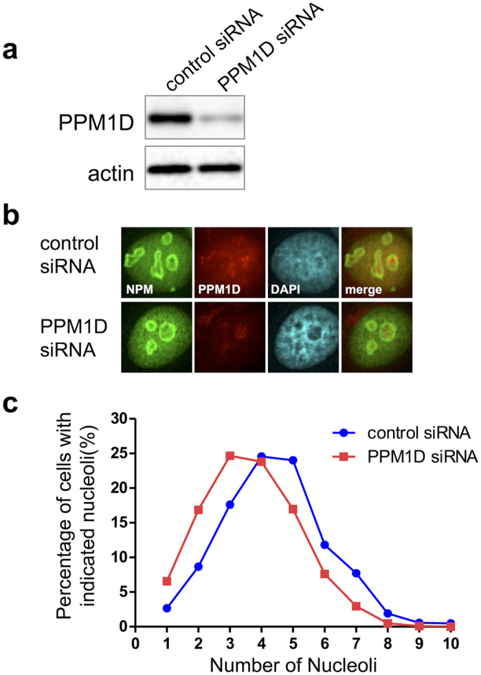
Figure 1. Nucleolar number was decreased by PPM1D knockdown.

MCF-7 cells were treated with PPM1D siRNA or control siRNA for 48 h, and were solubilized with 1x sample buffer for (a) Western blotting or (b) Immunocytochemistry. (a) PPM1D proteins were analysed by using Western blotting with rabbit polyclonal anti-PPM1D. (b) The subcellular localizations of NPM and PPM1D. PPM1D siRNA- or control siRNA-treated MCF-7 cells were fixed and stained with rabbit polyclonal anti-PPM1D and mouse monoclonal anti-NPM. (c) The percentage of cells with indicated nucleoli in PPM1D siRNA- or control siRNA-treated MCF-7 cells are shown. For quantitative analysis of the nucleolar number, stained-NPM was used as an indicator of the nucleoli. Number of cells used in the analysis are shown in Table 1; from at least two independent experiments.
