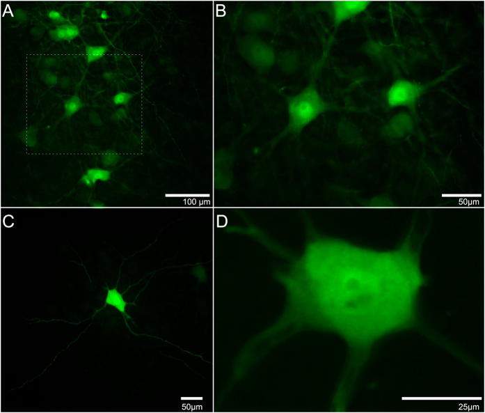
Figure 1. Representative images of enhanced green fluorescent protein (eGFP)-expressing motor neurons after intramuscular injections of Ad.eGFP at the motor end plate (MEP) region in triceps brachii and obtained from an adult mouse seven days after injection.
(A) Photomicrograph of a horizontal section through the ventral horn of the 5th cervical spinal cord segment (C5) showing typical eGFP expression in motor neurons. (B) Magnification of (A). (C) A single motor neuron within the spinal cord to show its axon and dendritic arborisation. (D) A z-stack of a different motor neuron at higher magnification. Both (C,D) were obtained from an adult mouse three days after intramuscular injection.

