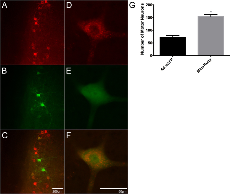
Figure 3. Representative images of Ad.eGFP and Mini-Ruby-positive motor neurons in the 7th cervical spinal cord segment (C7), seven days after 40 μl intramuscular injections of the cocktail at the MEP region in triceps brachii.
The TRITC and the FITC channels allow for the visualisation of Mini-Ruby (A) and eGFP (B), respectively. (C) Overlay of the two channels. Scale bar = 100 μm. (D–F) Higher magnification of a double-positive eGFP expressing and Mini-Ruby labelled motor neuron from a different section to that found in (A–C). Scale bar = 50 μm. (G) Quantitative analysis comparing the number of Ad.eGFP-transduced and Mini-Ruby-labelled motor neurons after intramuscular injections of a cocktail containing both constituents. T-test analysis reveals a statistical difference between the two groups (t = 7.89; df = 4; p = 0.0014). The error bars reflect the mean with SEM. Note, all cocktail-injected data were obtained from mice seven weeks of age and tissue was obtained seven days later.

