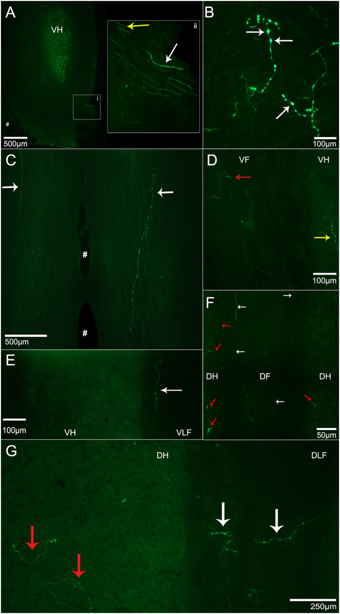
Figure 5. Representative images of eGFP-expressing axonal and dendritic processes extending from transduced motor neurons after Ad.eGFP injections were performed into the motor end plates of triceps brachii.

(A) Longitudinal sections through the ventral horn (VH) of the cervical spinal cord showing eGFP-expressing axons from triceps brachii transduced motor neurons extending through the ventrolateral funiculus (VLF) to exit via ventral roots. i) Axons of transduced motor neurons extending into the ventral root. ii) A close-up of i). White arrows indicate axons expressing eGFP extending into the ventral roots whereas the yellow arrows indicate the eGFP-expressing axons extending through the white matter located in the VLF. (B) eGFP-expression in processes located locally in VH grey matter. White arrows are suggestive of axonal boutons. (C) Longitudinal section of the white matter ventral to the VH in the cervical spinal cord after bilateral Ad.eGFP. White arrows indicate eGFP-expressing dendritic processes running through two cervical segments of the VLF. (D) Cervical section showing the VH and the ventral funiculus (VF). Red arrow indicates eGFP-expressing fibres located within the VF, whereas the yellow arrow indicates eGFP-expression located in the medial aspect of the VH. (E) Longitudinal section that includes the right VH and VLF. The white arrow points to one eGFP-expressing process located in the VLF. (F) Longitudinal section through the cervical spinal cord showing eGFP-expression in processes extending into the dorsal funiculus (DF). eGFP-expressing processes are extending along the rostro-caudal axis through the DF as indicated by white arrows and through the dorsal horn grey matter into the DF as indicated by red arrows. (G) eGFP-expressing processes extending into the DH and dorsolateral funiculus (DLF). White arrows indicate eGFP-expressing fibres extending into the DLF whereas the red arrows indicate eGFP-expressing processes extending into the DH. Images were obtained from mice from a variety of timepoints ranging from days 3–11. VH: ventral horn, VLF: ventrolateral funiculus, VF: ventral funiculus, DH: dorsal horn, DF: dorsal funiculus, #: the ventral median fissure.
