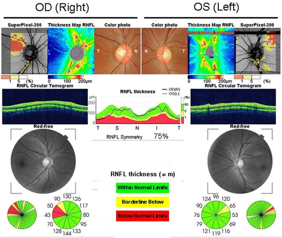
Fig. 4.

OCT shows that the right peripapillary retinal nerve fiber layer (pRNFL) is atrophic in the temporal quadrant, and the left pRNFL is thinning in the temporal superior quadrants. Green represents pRNFL thickness, which is within normal limits; yellow represents pRNFL thickness, which is below borderline; red represents pRNFL thickness, which is below normal limits
