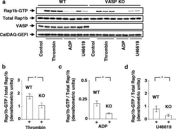
Fig. 1.

Agonist-induced Rap1b activation is reduced in VASP-null platelets. a Platelets (2 x10⁸) from wild type (WT) or VASP-null platelets (VASP KO) were unstimulated (control) or stimulated with thrombin (0.01 U/ml, 30s), ADP (10 μM, 1 min), or U46619 (1 μM, 1 min) (non-aggregating conditions). Thereafter, platelets were lysed and GST-RalGDS-RBD pull-down assays were performed as described in Methods. Proteins bound to GST-RalGDS-RBD were separated by 12 % SDS-PAGE, transferred to PVDF membranes, which were subjected to immunoblotting with anti-Rap1b Abs. The insets show representative Western Blots. Levels of Rap1b, VASP, or CalDAG-GEFI in the whole lysates used for the Rap1b pull-down assays were measured by Western blot analysis using appropriate Abs. The diagrams (b-d) illustrate densitometric analysis of the relative activities of Rap1b. Levels of Rap1b-GTP/total Rap1b in WT and VASP KO platelets stimulated with thrombin (b) (n = 5), ADP (c) (n = 3), or U46619 (d) (n = 3) were quantified by densitometry analysis using ImageJ software. *P < 0.05
