
Fig. 6.
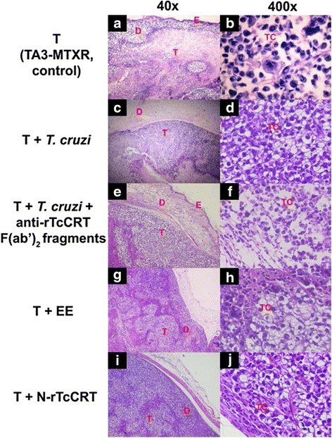
Mammary tumors from animals treated with either T. cruzi infection, EE or N-rTcCRT, display less aggressive histological patterns. TA3-MTXR tumor (T) extracted from representative animals: a b Non infected, control group; c d T. cruzi infected; e f T. cruzi- infected, inoculated with anti-rTcCRT F(ab’)2 fragments; g h Inoculated with an EE; i j Inoculated with N-rTcCRT. The following parameters were analyzed: E: Epidermis; D: Dermis layer; T: Tumor tissue (a, c, e, g, and i) and TC: Tumor cells (b, d, f, h and j)