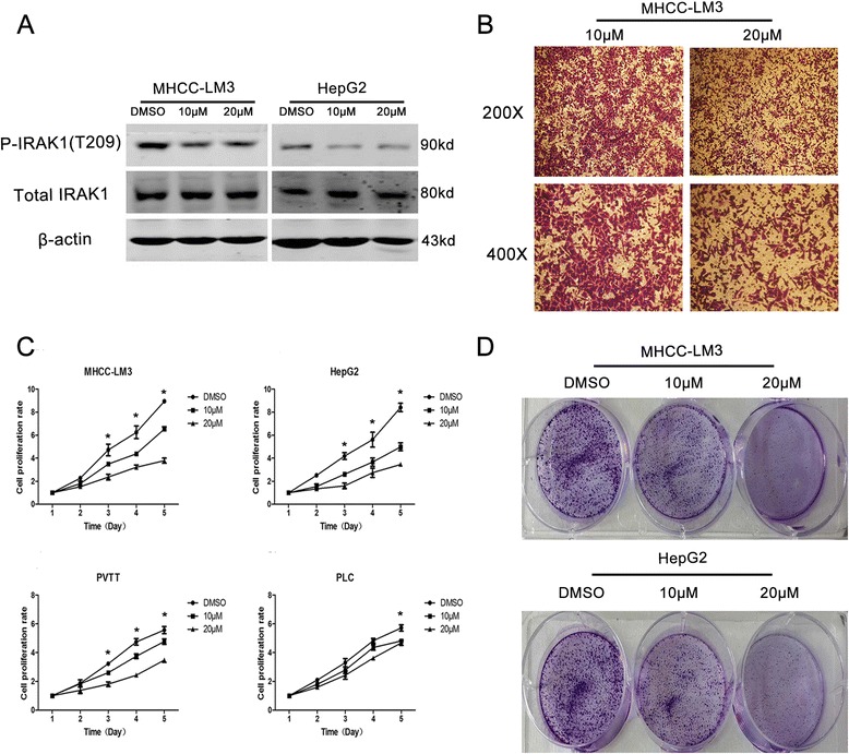
Fig. 4.

The inhibition of p-IRAK1 attenuated proliferation in HCC cells. a The protein levels of p-IRAK1 and total IRAK1 in SMMU-7721 and HepG2 with IRAK1/4 inhibitors (0, 10 μM, 20 μM). b Cell migration analysis in SMMU-7721 cell lines with IRAK1/4 inhibitors (0, 20 μM) treatment for 24 h. c The proliferation analysis in four different cell lines with IRAK1/4 inhibitors (0, 10 μM, 20 μM) for 1–5 days. d Colony formation analysis in SMMU-7721 and HepG2 cells with IRAK1/4 inhibitor (0, 10 μM and 20 μM) for 48 h
