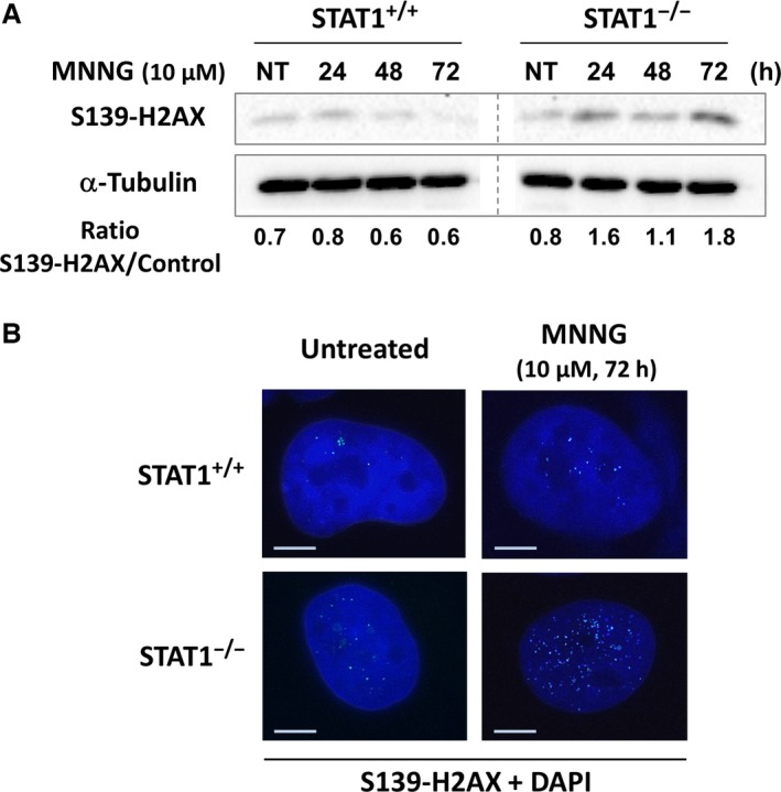
Figure 2.

Higher activation of histone H2AX following MNNG exposure in the absence of STAT1. STAT1+/+ and STAT1−/− cells were treated for 1 h or not (NT) with 10 μM MNNG. (A) Western blot analysis of S139‐H2AX at the indicated time‐points. A typical experiment out of three is shown. α‐Tubulin was used as loading control. Both panels are cut from the same western blot. (B) 72 hrs following treatment, cells were also fixed and immunostained for S139‐H2AX. Nuclei were counterstained with DAPI. A typical immunostaining out of two is shown. Scale bars, 5 μm.
