
Figure 7.
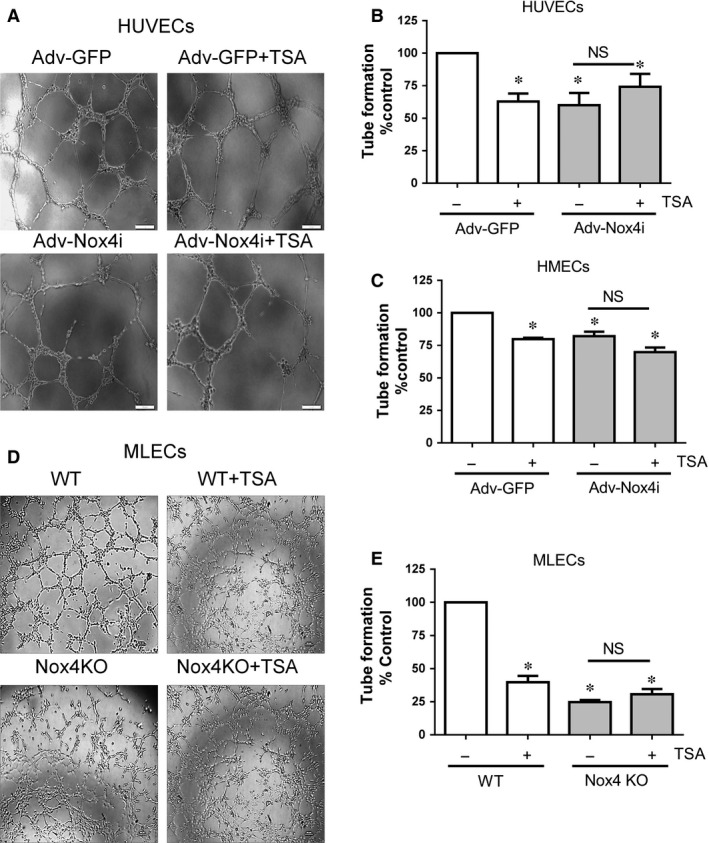
Effect of TSA on endothelial cell capillary formation. HUVECs and HMECs were infected with either Adv‐GFP or Adv‐Nox4i for 48 hrs. HUVECs HMECs and MLECs formed capillaries on growth factor reduced Matrigel® at 8 hrs. (A) Representative high magnification images of a tube formation in (A) HUVECs and (D) MLECs (scale bar represents 20 μm). (B) HUVEC and (C) HMEC cells were treated at the cell seeding on Matrigel® with tricostatin A (TSA; 0.33 μM) for 8 hrs. (C) Similarly, wild type (WT) MLECs and Nox4 knockout (Nox4 KO) MLECs were also treated with TSA (0.33 μM) for 8 hrs. Tube length was measured using ImageJ software and expressed as percentage control. Data expressed as mean ± S.E.M. from 4 to 6 independent experiments. *P < 0.05 from Adv‐GFP or WT without TSA treatment. NS: Not significant.