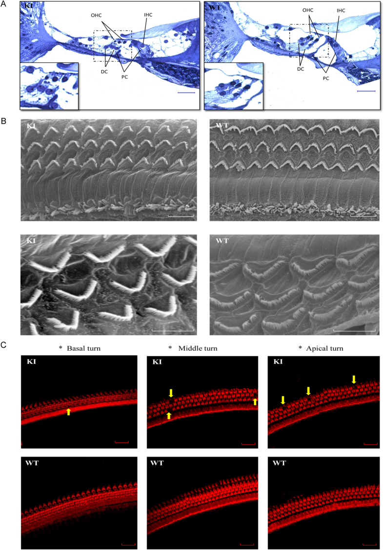
Figure 2. Cochlea morphology of the homozygous p.V37I knock-in (KI) and wild-type (WT) mice at age one year.
(A) Semithin histology sections showing the normal gross morphology of the KI mouse cochleae. Scale bars: 100 μm. (B) Scanning electron microscopy showing normal cellular structure and arrangement of the hair cell bundles of the KI mice. Scale bars: 10 μm (top) and 5 μm (bottom). (C) Confocal immunofluorescence microscopy showing minor loss of the outer hair cells (arrows) at the apical, middle and basal turns of the KI mouse cochleae. F-actin was stained with rhodamine phalloidin (1:100) to identify the hair cells. Scale bars: 20 μm.

