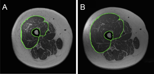
Figure 1.

A representative T1-weighted MRI of the mid-thigh (A) pre-training and (B) post-training in an individual with complete spinal cord injury.
The knee extensor muscle group is traced out to highlight the extensive skeletal muscle hypertrophy following 16 weeks of resistance training. Bony and soft tissue landmarks were used to match pre-training and post-training images to ensure the exact anatomical location.
