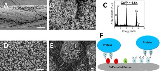
Figure 1.

Calcium phosphate (CaP) coatings can be grown on a variety of materials in modified simulated body fluids (mSBF).
(A) Scanning electron microscopy micrographs of CaP coated Vicryl sutures show a continuous layer of CaP coating. (B) Higher magnification shows the nanoporous plate like structure creating a large surface area for protein binding. (C) Energy dispersive spectroscopygraph obtained from a CaP coated suture reveals peaks of calcium and phosphate crystals. (D) Scanning electron microscopy micrographs of CaP coated β-tricalcium phosphate microparticles shows a continuous layer of CaP coating and (E) higher magnification reveals a nano-porous structure similar to the CaP coated sutures. (F) The CaP coatings are a mosaic of positively charged crystal calcium ions and clusters of negatively charged oxygen atoms associated with crystal phosphates and the majority of protein binding to the CaP coating is a combination of metal affinity and phosphoryl cation exchange. The illustration shows proteins binding via metal chelation between carboxyl clusters and crystal calcium ions and proteins binding via cation exchange between negatively charged oxygen atoms associated with crystal phosphates and positive amines on the protein. Scale bars: A and D: 100 μm, B and E: 1 μm.
