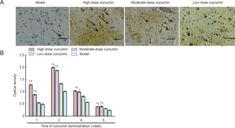
Figure 4.
S100 immunoreactivity in crosssections of L4–6 spinal cord segments 8 weeks after curcumin administration.
(A) Immunohistochemical staining of S100 protein 2 weeks following curcumin administration at 40× magnification. Scale bar: 200 μm. Arrows indicate positive immunoreactivity for S100, which is seen as fine brown particles in the cytoplasm. (B) Staining intensity. The staining intensity in the high- and moderate-dose curcumin groups is stronger than that in the low-dose curcumin and model groups. Data are expressed as the mean ± SD (n = 5). One-way analysis of variance and Dunnett's tests were used to analyze the difference among groups. *P < 0.05, vs. model group; #P < 0.05, vs. low-dose curcumin group. High-, moderate-, low-dose curcumin groups: curcumin 40, 20, 10 mg/kg/d, respectively.

