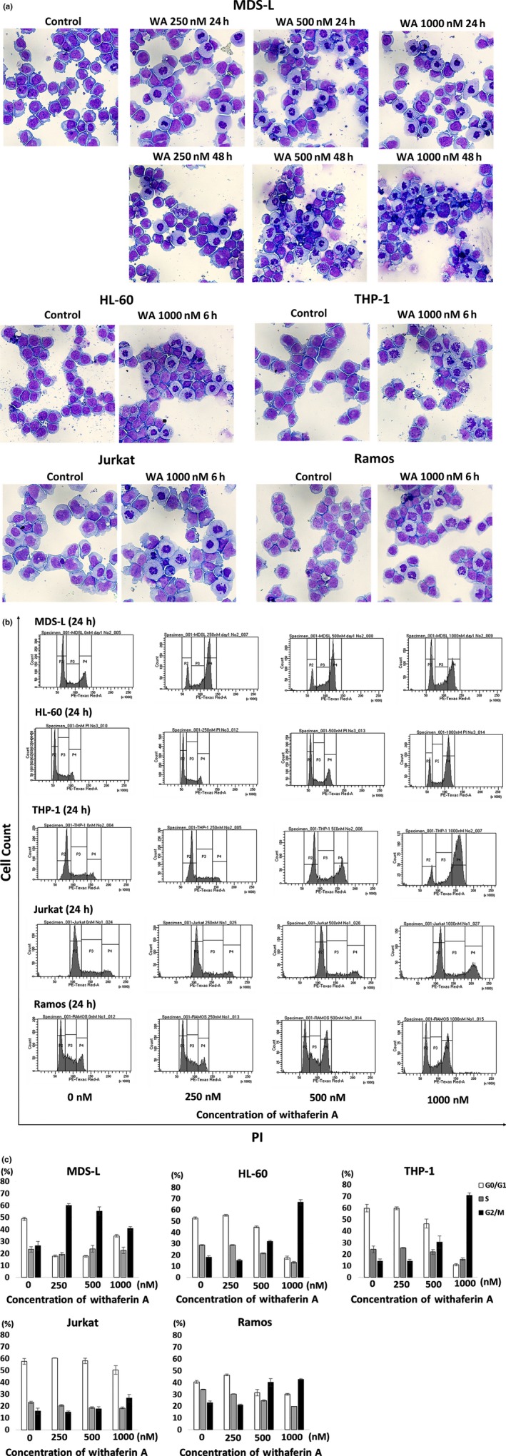
Figure 2.

Effects of withaferin A (WA) on the morphology and cell cycle of myelodysplasia and leukemia cell lines. (a) MDS‐L cells were cultured without treatment (control) or treated with 250, 500 and 1000 nM WA for 24 and 48 h. HL60, THP‐1, Jurkat and Ramos cells were cultured without treatment (control) or treated with 1000 nM WA for 6 h. Cytospin preparations were stained by May‐Gruenwald‐Giemsa (original magnification x400). Representative images by each treatment are shown. (b) The cell cycle analyses by propidium iodide (PI) staining are shown. MDS‐L, HL‐60, THP‐1, Jurkat and Ramos cells were treated with indicated concentrations of WA for 24 h, and the cells were stained with PI and analyzed by flow cytometry. Representative histogram patterns by each treatment are shown. (c) The cell fractions at G1, S and G2⁄M phase are presented by white, gray and black bars, respectively. The data represent the mean values with SD from three independent experiments.
