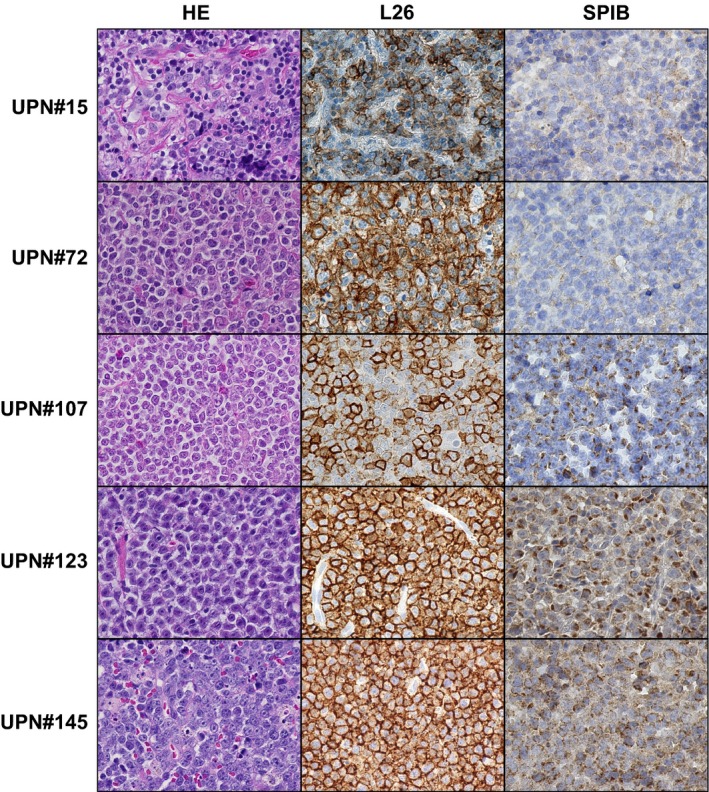
Figure 1.

SPIB immunostaining of DLBCL specimens. Representative images of DLBCL stained by HE (left panels), anti‐CD20 antibody, L26 (center panels) and SPIB (right panels) are shown. CD20 positive tumor cells diffusely proliferate in the lymph node. Specimens UPN#15 and UPN#72 were assessed as SPIB negative, while UPN#107, UPN#123 and UPN#145 were assessed as SPIB positive (original magnification 400 × , Keyence BZ‐9000).
