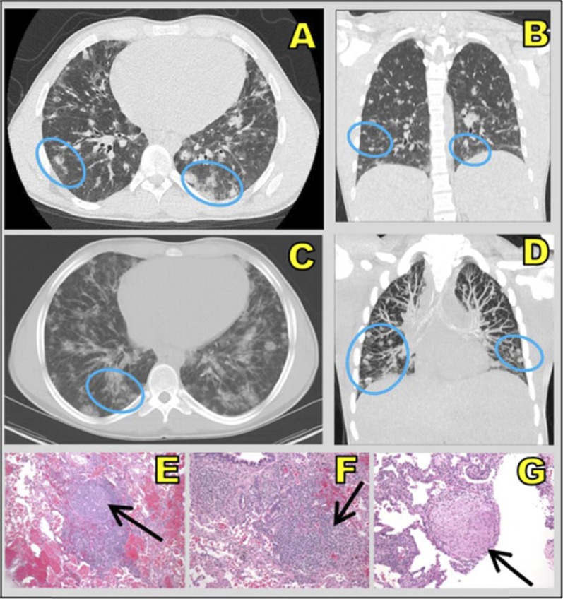
Figure 1.
(A and C) Axial and (B and D) coronal computed tomographic scans show ill-defined, diffuse ground-glass nodules (blue ovals), which are common in fungal infection, in the lower lobes. Right lung biopsy (hematoxylin and eosin stain; original magnification, ×10) shows (E) a nodular lymphoid infiltrate with a prominent “naked” reactive germinal center, (F) a peribronchial lymphocytic infiltrate, and (G) a nonnecrotizing granuloma composed of epithelioid and rare multinucleated giant cells (black arrows) associated with interstitial lymphocytic infiltrate. This patient (patient 6 from Table 2) denied pulmonary symptoms and is an active hockey player.

