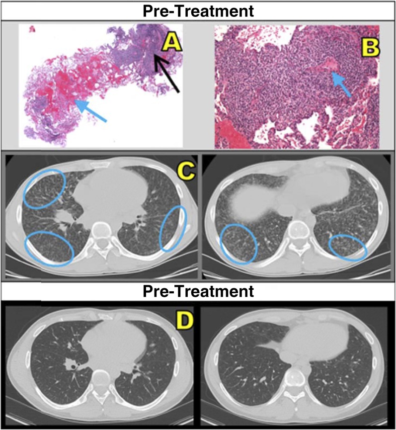
Figure 2.
(A) Core lung biopsy (hematoxylin and eosin stain; original magnification, ×2) shows intraalveolar hemorrhage (blue arrow) and patchy peribronchial/perivascular lymphocytic infiltrate (black arrow). (B) Dense lymphocytic infiltrate and interstitial widening, sparing blood vessels (blue arrow) (original magnification, ×10). (C) Pretreatment computed tomographic scans show miliary-like, small, ill-defined, diffuse micronodular ground-glass nodules (blue ovals) in right middle and bilateral lower lobes. (D) This patient (patient 4 from Table 2) showed interval improvement after treatment with mycophenolate mofetil and high-dose methylprednisolone.

