Figure 4.
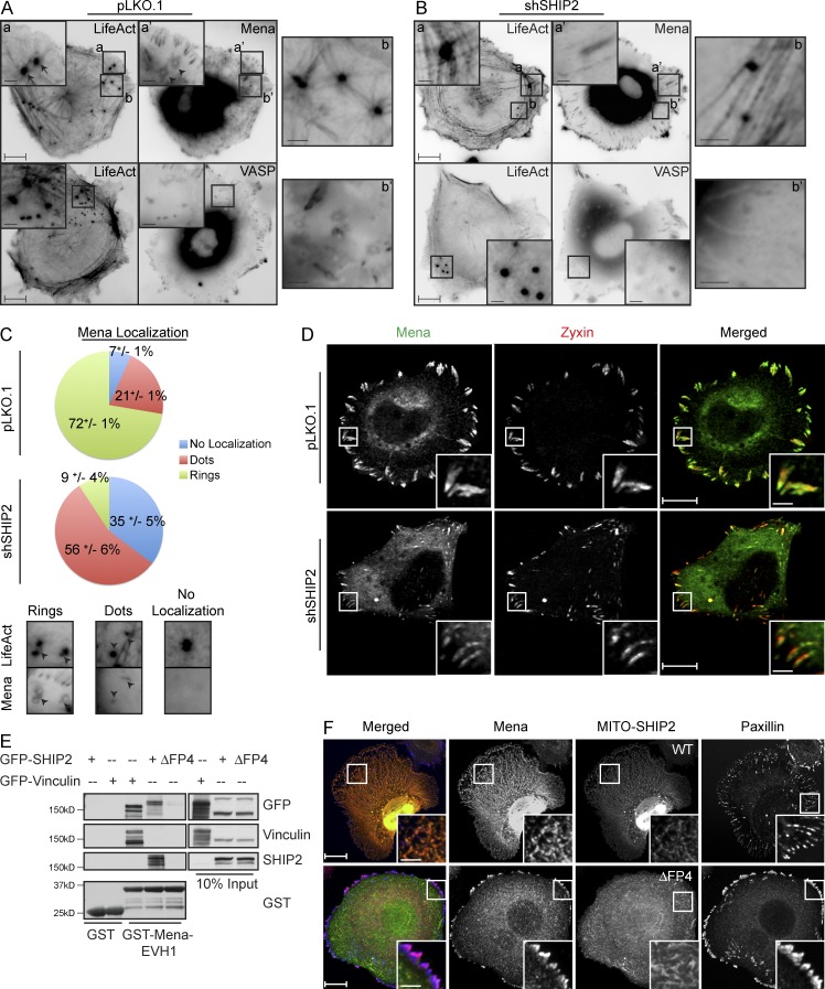
SHIP2 recruits Mena, but not VASP, to promote proteolytically active invadopodia formation. (A) MDA-MB-231-pLKO.1 cells were transiently transfected with tagRFP-LifeAct and GFP-Mena or GFP-VASP and subjected to time-lapse video microscopy. Single frame illustrates the differential localization of Mena and VASP with respect to filamentous actin at invadopodia protrusions. (B) As in A, MDA-MB-231-shSHIP2 cells were transiently transfected with tagRFP-LifeAct and GFP-Mena or GFP-VASP and subjected to time-lapse video microscopy. Single frame illustrates that, under SHIP2-depleted conditions, Mena localizes to focal adhesions but not to filamentous actin at invadopodia protrusions, whereas VASP still localizes to filamentous actin at invadopodia protrusions. (C) GFP-Mena and tagRFP-LifeAct were transiently coexpressed in SHIP2-depleted MDA-MB-231 cells and subjected to the time-lapse imaging. Three different localization patterns of Mena relative to F-actin punctae (tagRFP-LifeAct) are observed. The relative distribution of Mena localization pattern under SHIP2-depleted condition compared with control is quantified and depicted as a pie chart. Quantification addresses percentage of invadopodia from 36 cells during the course of the video across three independent experiments. (D) MDA-MB-231-pLKO.1 and MDA-MB-231-shSHIP2 cells were immunostained with Zyxin and Mena, and confocal images were acquired at the ventral surface. (E) GST and GST-Mena-EVH1 were coupled to glutathione-Sepharose beads and used to pull down interaction partners from the lysates of HEK293 cells transiently transfected with GFP-Vinculin, GFP-SHIP2, or GFP-SHIP2ΔFP4. Resulting samples were probed for GFP, Vinculin, and SHIP2 as indicated. (F) Mitochondria-targeted SHIP2 (Mito-SHIP2) or Mito-SHIP2ΔFP4 was transiently coexpressed together with mCherry-Mena in SKBr3 cells and immunostained as indicated to establish the localization of Mena with respect to SHIP2 and focal adhesions. All quantified data indicate the mean values ± SE from at least three independent experiments. Bars: 10 µm; (inset) 2 µm.

