
Figure 3.
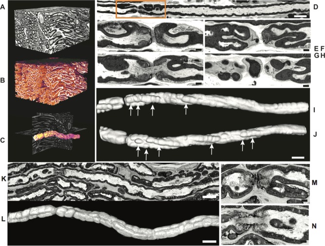
Serial blockface scanning EM analysis of nuc‐ErbB3 KI sciatic nerves shows aberrant myelination. (A–J) Three‐dimensional reconstruction of image stack (120 × 80 × 60 μm). Intensity‐based segmentation of myelinated fibers identified many with irregular myelin foldings (B, false color scale). Myelin foldings at nodes are elaborate as illustrated for a representative axon from this set (C, shown in orange on orthogonal slices). A single slice view (D) indicates myelin outfoldings are prominent around the node. In single slices (E–H, from box in D), myelin folds appear isolated, but in 3D view of the compact myelin (I, same orientation as D and J rotated through 180°) aberrant myelin foldings were continuous along the internode and not restricted to the nodal region (arrows). (K–N) Single slice view of control (WT) sciatic nerves shows that the nodal axoplasm is open and continuous and the perinodal crenulation and myelin outpocketing is not as pronounced as in nuc‐ErbB3 nerves. Three‐dimensional view of the compact myelin along the axon shows lack of aberrant myelin foldings along the axon (L). Scale bars A–D, K, 5 μm; E–J, L–N 1 μm). [Color figure can be viewed in the online issue, which is available at wileyonlinelibrary.com.]